
- Education
-
Research
Current research
Talent
-
Collaboration
Businesses
Government agencies and institutions
Alumni
-
About AU
Organisation
Job at AU
The faculty’s new bioimaging core facility is now opening for all Health researchers who wish to make use of the eight advanced microscope systems which are currently available. The microscopes are installed in the Skou Building at the bottom of the University Park.
2019.09.03 |
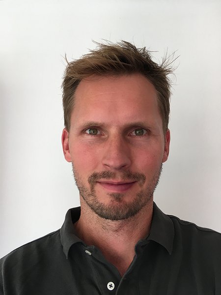
The microscopes will provide many researchers with exciting opportunities, but new users should be properly trained before being able to use the equipment by themselves, because it’s one thing to take images with the equipment, but something else entirely to analyse and interpret the results correctly, Christian Garm explains. Photo: AU
After years of planning and with researchers longing to begin using it, the new Bioimaging Core Facility at the Department of Biomedicine is finally ready. As the first of its kind in Aarhus, the facility brings together this type of advanced microscope systems for the benefit of everyone who carries out research in the life science field, explains MSc and PhD Christian Garm who has been working as head of the Imaging Core Facility since earlier this summer. Christian has previously worked as both a researcher and microscope specialist at Olympus, who have developed the majority of the equipment at the facility.
“The various microscopes will provide many of the faculty’s, and for that sake the university’s other researchers, with exciting opportunities, though this is not the type of equipment that you just walk in and use. New users should be properly trained before being able to use the equipment by themselves, because it’s one thing to take images with the equipment, but something else entirely to analyse and interpret the results correctly,” says Christian Garm. He estimates that the value of the eight microscopes available so far is around DKK 13 million.
The facility comprises, among other things, a multiphoton microscope for live imaging of animals – a microscope that makes it possible to look into the brain of a mouse or rat while it is alive. When it comes to the other microscope systems, Christian Garm highlights a new slide scanning system for high quality fluorescence and bright field scanning, together with three confocal microscopes including a new LSM800 with Airyscan from Zeiss.
All the equipment is described in detail on the new website http://imaging.au.dk/, which is also where you can register as a user and sign up for an introductory course with Christian Garm.
He explains that all the microscope systems are installed in the Skou Building, with one consequence being that researchers from e.g. Skejby must actually visit the location in the University Park to use the equipment.
“The equipment is complex and it’s not something you just move around, so it’s much easier to invite researchers to come to the Skou Building. However, I’m working to allow researchers to access imaging processing software from their own computer and this will soon be possible,” says Christian Garm. His best guess is that ninety per cent of the core facility’s users will come from the faculty, while the other ten per cent will be external customers.
He goes on to explain that the eight systems cover a very broad spectrum of techniques within microscopy and that there has been technological progress on two fronts in particular which can elevate the facility to an even higher level – Lattice SIM (Structured Illumination Microscopy) and light sheet microscopy.
“The first technique enables super resolution, and with the current systems we can separate molecules that are separated with approximately 120 nm, though we dream of squeezing this all the way down to 20-30 nm,” he explains.
”Light sheet microscopy is a technique in which complete organs such as brains are made fully transparent, making it possible to then use special lighting across the sample to create perfect 3D representations throughout the sample,” he adds. Funding is being sought for the two technologies on an ongoing basis.
Users from the university pay a modest user fee and prices can be seen on the website. External customers are charged more.
The core facility is funded by contributions from the faculty, the department, various foundations and research groups at the department. Christian Garm points out that it has only been possible to establish the facility due to the good cooperation between the department and a number of group leaders, who have been willing to share existing expensive equipment.
Morten Schallburg Nielsen is the academic head of the facility.
PhD and Manager Christian Garm
Head of Bioimaging Core Facility
Skou Building (1116), Høegh-Guldbergs Gade 10
Email: christian.garm@biomed.au.dk
Mobile: (+45) 9352 1888