- Education
-
Research
Current research
Talent
-
Collaboration
Businesses
Government agencies and institutions
Alumni
-
About AU
Organisation
Job at AU
A group of researchers at Aarhus University have gained unique access to brain tissue that is left over following operations. As the first in Denmark, they can now test new drugs or study the body's own neurotransmitters on living pieces of human brain in the laboratory.
2021.07.05 |

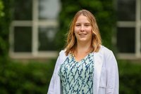
For the first time in Denmark, researchers have had the opportunity to use living human brain tissue for preclinical studies. The ground-breaking research has emerged in a collaboration between the Capogna Laboratory at the Department of Biomedicine at Aarhus University and the neurosurgery team at Aarhus University Hospital. Post.doc Emma Louise Louth has headed the first study. Photo: Simon Byrial Fischel
When patients have a tumour in the brain removed at Aarhus University Hospital, Postdoc Emma Louise Louth waits in the operating theatre. She is there to fetch a small piece of living brain tissue.
The researcher from Aarhus University has brought a container with artificial cerebral spinal fluid – an imitation of the substance that protects the brain and spinal cord. The liquid is oxidised and ice-cold, and it can keep the small piece of brain alive in the back seat of Emma Louise Louth's car until she arrives at the laboratory in the University Park.
Once there, she immediately cuts the little piece of human brain into thin slices and places it in a container with ions and salts at room temperature. Here she can keep it alive for 12 hours.
During this time, Emma Louise Louth and the other members of the research group headed by Professor Marco Capogna have the chance to carry out studies and experiments that have previously only been possible using laboratory animals.
The group's first study on human brain tissue was published in the spring of 2021 in the international journal Frontiers in Cellular Neuroscience, and the research group has compared the dopamine-enhanced connections between neurons in humans and mice.
"To borrow an analogy from another researcher: Mouse studies versus human studies are basically like looking at a Nokia 3310 when trying to repair an iPhone. They have the same basic functions – but there is much greater complexity in the human brain. We even know that there are differences in the types of cells and the expression of certain receptors. Therefore, being able to test directly in human tissue is a unique opportunity," explains Emma Louise Louth.
She has headed the first study and this shows that dopamine – which is known for its role in the brain's reward system – strengthens the connections between neurons in the human brain. This is important knowledge and can lead to new treatment opportunities, for example in connection with rehabilitation after a stroke or other types of acute brain damage, where patients lose synaptic connections in the brain and need to form new ones.
"We’ve been given the opportunity to show that dopamine plays a different role in humans and mice. This is a really good example of how the effect of a drug or a neurotransmitter varies between species, and it highlights the importance of being able to test drugs directly on human tissue," says Emma Louise Louth.
A very special collaboration between the neurosurgeons at Aarhus University Hospital and professor Capognas’ research group at the Department of Biomedicine has made it possible for the first Danish research into living human brain.
When patients with a brain tumour arrive at a consultation before an operation, they are informed that the surgeons will have to remove a certain amount of healthy brain to get at the tumour. They are then asked whether they wish to donate this piece of brain tissue to a research project. Virtually everyone says yes, explains Emma Louise Louth:
“People are very happy to donate to something useful.”
Therefore, roughly every other week she drives to the hospital in Skejby to collect fresh and healthy tissue. She does not know who the brain tissue belongs to – the research group only receives basic information about which area of the brain it comes from, how far it is from the tumour, and about the patient's gender, age group, medicine consumption and any other diseases.
The ground-breaking research is only possible because the neurosurgeons from Jens Christian Hedemann Sørensen's team at Aarhus University Hospital, together with Professor Anders Rosendal Korshøj from the Department of Clinical Medicine, are willing to make an extra effort. Researchers and surgeons have held many meetings about the project, and the surgeons have a list of golden rules that they must follow during the operation.
"For the surgeons, it’s a completely different process than normal. They have to be very careful with the scalpel, and they should preferably not use the coagulator, because it generates heat and that’s not good for my living tissue. Nor should they pinch too hard with the tweezers when removing the tissue. Each surgeon supplying tissue must be involved, and that requires a lot of communication," she explains.
"Fortunately, we're dealing with a group of inquisitive and interested neurosurgeons who wish to interact with the research side of things."
Certain philosophical questions are unavoidable when a living piece of the most central organ in the human nervous system lies there in a petri dish – and is alive. Can it feel pain? Can it think?
No, it can’t, says Emma Louise Louth:
"Every emotion or thought must go through many parts of the brain. The piece we work on is the size of the outermost part of your thumb, and it’s no longer connected to other areas of the brain. I understand why people wonder whether the neurons in the petri dish have a memory, but it’s simply not possible."
The research group is currently working on a method that can keep the small slices of brains alive for up to ten days.
The group has begun collaborating with the Allen Institute for Brain Science in the USA, which produces fast-acting viruses. The researchers hope to be able to use the virus to make specific neurons in the brain luminous, and thus learn more about how they communicate.
"The first project was primarily about establishing the cooperation with the surgeons and gaining access to healthy, living tissue. Now we're planning all the exciting studies we can do with it," says Emma Louise Louth.
The researcher cuts the brain tissue into thin slices and places a piece under a microscope.
The researcher then pierces the brain tissue with a glass pipette with an electrode in the tip. In this way, the researcher can activate each neuron – and measure and record the electrical signals flowing through it. The technique is called whole cell electrophysiology.
This means that the researcher can test the neurons' reaction to various substances, such as e.g. dopamine. She can see how cells react to the substance, how neurons communicate, and how the connections change over time.
Postdoc Emma Louise Louth
Department of Biomedicine, Aarhus University
Email: emma.louth@biomed.au.dk
Phone: +45 81923938